What Is Giant Cell Arteritis?
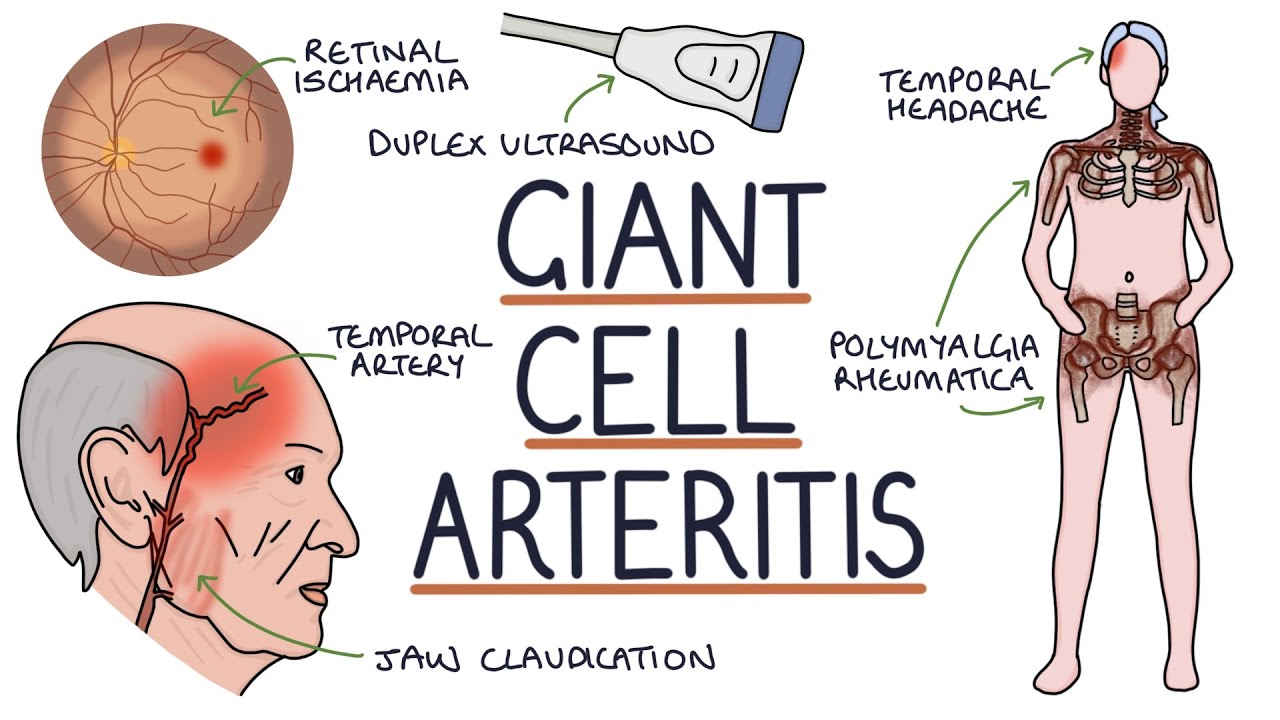
Giant cell arteritis is an inflammatory condition that affects medium and large blood vessels, most often in adults over age 50. It commonly involves arteries in the head, scalp, and temple area. Because these blood vessels help supply blood to important structures, including the eyes, GCA is treated as an urgent medical concern.
Patients with suspected GCA may experience symptoms such as:
- New or unusual headache
- Tenderness near the temples
- Scalp tenderness, especially when brushing hair
- Jaw pain or tiredness while chewing
- Blurred vision
- Double vision
- Temporary vision dimming or blackouts
- Sudden vision loss
- Fatigue, fever, or unexplained weight loss
- Shoulder, hip, or muscle stiffness
Why Would I Need a Temporal Artery Biopsy?
A temporal artery biopsy may be recommended when your symptoms, bloodwork, or eye exam findings suggest giant cell arteritis. Blood tests such as ESR and CRP can indicate inflammation, but they do not always identify the exact cause. A biopsy allows a small sample of the temporal artery to be examined under a microscope for signs of inflammation.
This procedure may be especially important if you have symptoms such as a new headache, temple pain, jaw pain with chewing, or vision changes. For patients in Lubbock, West Texas, and the surrounding region, having access to an oculoplastic surgeon who performs temporal artery biopsy can help support faster diagnosis and coordinated care.
In some cases, treatment for suspected GCA may begin before biopsy results are available because the risk to vision can be serious. Your physician will explain the timing of treatment and biopsy based on your symptoms and medical history.
How Giant Cell Arteritis Can Threaten Vision
The eye depends on steady blood flow to keep the optic nerve and retina healthy. When giant cell arteritis causes inflammation in blood vessels, blood flow can become restricted. If the optic nerve does not receive enough oxygen-rich blood, sudden vision loss can occur.
This vision loss is often painless and may happen quickly. In some cases, one eye is affected first, and the other eye may also be at risk. That is why GCA is not something to “watch and wait” on. Prompt evaluation, treatment, and diagnostic testing are essential.
What Happens During a Temporal Artery Biopsy?
A temporal artery biopsy is typically performed as an outpatient procedure using local anesthesia. The area near the temple is cleaned and numbed to keep you comfortable during the biopsy.
During the procedure, Dr. Ray makes a small incision near the temple or hairline area. A small section of the temporal artery is carefully removed and sent to a pathology lab for review. The incision is then closed with careful attention to healing and appearance.
Most patients return home the same day. Your care team will provide detailed instructions for caring for the incision site after the procedure.
What to Expect After the Procedure
After a temporal artery biopsy, it is normal to have some mild tenderness, swelling, bruising, or soreness near the incision. These symptoms are usually temporary.
Your post-procedure instructions may include:
- Keeping the incision clean and dry
- Avoiding strenuous activity for a short period
- Taking medications as directed
- Returning for follow-up care or suture removal, if needed
- Watching for signs of infection, such as increasing redness, warmth, drainage, or worsening pain
A small scar may remain, but the incision is usually placed to minimize visibility when possible.