Choroidal Nevus: Signs, Risks, and When to See a Retina Specialist
A choroidal nevus is a flat, mole-like spot inside the eye. Most are benign and never cause trouble, but a small percentage can change over time. At West Texas Eye Associates, our retina team, including Douglas Jin, MD, evaluates, documents, and monitors choroidal nevi so you understand your risk and know exactly when treatment is necessary.
What Is a Choroidal Nevus?
A choroidal nevus is a pigmented lesion in the choroid (the vascular layer beneath the retina). It is usually discovered during a dilated eye exam and looks gray-brown on retinal photos. Most people have no symptoms; occasionally, patients notice blurred vision, flashes/floaters, or a shadow if fluid develops under the retina. Authoritative clinical overviews note that nevi are common and typically stable, but they must be differentiated from early melanoma and other mimickers.
Why Monitoring Matters
Large data sets from tertiary centers show that a small fraction of nevi grow into melanoma over time; risk rises when specific features are present (see below). In long-term cohorts, cumulative transformation risk across many patients remains low but non-zero, reinforcing the need for periodic imaging and follow-up.
The Major Risk Features (TFSOM-DIM)
Specialists use the TFSOM-DIM mnemonic to flag higher-risk nevi:
Thickness >2 mm
Fluid (subretinal)
Symptoms (decreased vision, flashes/floaters)
Orange pigment (lipofuscin)
Margin near the optic disc
Diameter >5 mm
Imaging features (I): ultrasonographic Medium reflectivity/hollowness, autofluorescence changes, OCT abnormalities
These features correlate with growth risk and guide how closely we follow you or when we involve an ocular oncologist.
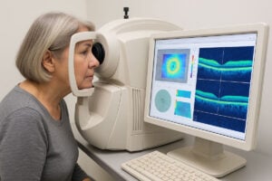
How We Evaluate and Track a Nevus
Dilated retinal exam & color fundus photography to document baseline size and borders
Optical Coherence Tomography (OCT) to detect subtle subretinal fluid or retinal changes
Fundus autofluorescence to look for orange pigment/lipofuscin
B-scan ultrasound to measure thickness and internal reflectivity
Risk stratification + schedule for follow-up (for many patients, 6–12 months; sooner if features change)
Modern imaging improves the detection of the risk features above and helps separate benign nevi from small melanomas.
When a Nevus Needs Treatment
Most nevi are observed only. If a lesion shows growth or high-risk features, we coordinate with ocular oncology to discuss options (e.g., plaque radiotherapy) appropriate for small choroidal melanoma. Patient-facing resources on ocular melanoma provide a useful overview of why early detection matters.
Symptoms You Shouldn’t Ignore
New or worsening blurred or distorted central vision
Flashes, an increase in floaters, or a dark curtain/shadow
Douglas Jin, MD, is a retina specialist in Lubbock who cares for patients with choroidal nevi and other retinal conditions. He uses modern retinal imaging (OCT, fundus autofluorescence, ultrasound) to document baseline size and watch for risk features, and he coordinates with ocular oncology if treatment is indicated.
Your Visit to West Texas Eye Associates
You’ll receive a clear, illustrated report with photographs and measurements, your personalized risk level, and a follow-up plan. We’ll tell you exactly what to watch for at home and when to return sooner than scheduled. Your evaluation may be with Douglas Jin, MD, our retina specialist in Lubbock.
Living With a Choroidal Nevus: Practical Tips
Keep annual dilated eye exams (or your personalized interval)
Tell your doctor about new visual symptoms right away
Bring previous photos/reports if you’re new to our practice
Protect eyes from UV and manage cardiovascular risk factors; while evidence is evolving, healthy habits support overall ocular health
FAQ
Is a choroidal nevus cancer?
No. It’s a benign “mole” inside the eye. A small percentage of individuals develop changes that suggest melanoma, which is why regular monitoring is crucial.
How often should I be checked?
It depends on size and features. Many patients are seen every 6–12 months; higher-risk nevi are followed more often.
What tests will I have?
Typically, retinal photos, OCT, autofluorescence, and ultrasound to measure thickness. These tests are painless and take just minutes.
What raises the risk that a nevus will grow?
Thickness over 2 mm, subretinal fluid, symptoms, orange pigment, margin near the optic disc, larger diameter, and certain ultrasound findings (TFSOM-DIM).
If it turns into melanoma, what are the treatments?
Options may include plaque radiotherapy or other modalities delivered by ocular oncology; your retina specialist will coordinate care. Patient resources on uveal melanoma are available from the National Eye Institute.
We Can Help, Today
Most choroidal nevi remain asymptomatic for life, but they still require expert evaluation and modern imaging. If you’ve been told you have a nevus—or you’re noticing new visual symptoms, schedule a comprehensive retina evaluation at West Texas Eye Associates. We’ll document a precise baseline, explain your risk, and set a follow-up plan tailored to you.
Related Posts
Schedule an appointment online
Book Your Next Appointment Entirely Online.
Find An Appointment That Works For You!
To provide the best experiences, we use technologies like cookies to store and/or access device information. Consenting to these technologies will allow us to process data such as browsing behavior or unique IDs on this site. Not consenting or withdrawing consent, may adversely affect certain features and functions.
Functional
Always active
The technical storage or access is strictly necessary for the legitimate purpose of enabling the use of a specific service explicitly requested by the subscriber or user, or for the sole purpose of carrying out the transmission of a communication over an electronic communications network.
Preferences
The technical storage or access is necessary for the legitimate purpose of storing preferences that are not requested by the subscriber or user.
Statistics
The technical storage or access that is used exclusively for statistical purposes.The technical storage or access that is used exclusively for anonymous statistical purposes. Without a subpoena, voluntary compliance on the part of your Internet Service Provider, or additional records from a third party, information stored or retrieved for this purpose alone cannot usually be used to identify you.
Marketing
The technical storage or access is required to create user profiles to send advertising, or to track the user on a website or across several websites for similar marketing purposes.